Hello
From the heart of Seomyeon,
drawing tomorrow's smile,
This is Smile Digital Dental Clinic 🙂
If a problem has appeared again around the roots of a tooth that previously had root canal treatment,
does the tooth have to be extracted?
Even for a tooth that has had root canal treatment, after checking the condition of the previous root canal filling, the roots, the remaining tooth structure,
and the surrounding periodontal tissue, there are cases in which we can consider keeping the natural tooth
through root canal retreatment.
On the other hand, if there is a crack or fracture in the root, or the tooth is in a condition that is difficult to restore again,
root canal retreatment may not be suitable.
Today we introduce the case of a man in his 40s living in Germany who visited because of a problem with a molar that had previously had root canal treatment. After root canal retreatment, we followed the tooth over a long period, at 6 months, 1 year, 2 years, 4 years, and at the most recent checkup on October 2, 2026.
Table of contents
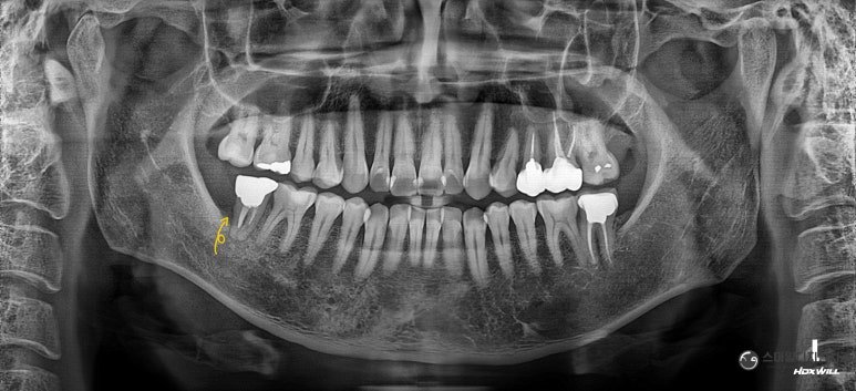
Panoramic X-ray before treatment
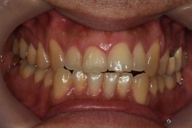
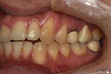
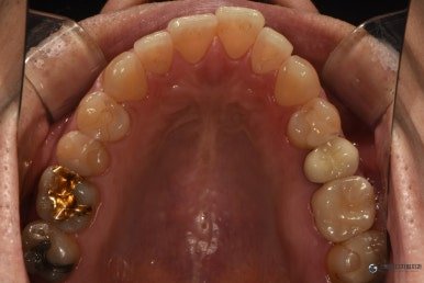
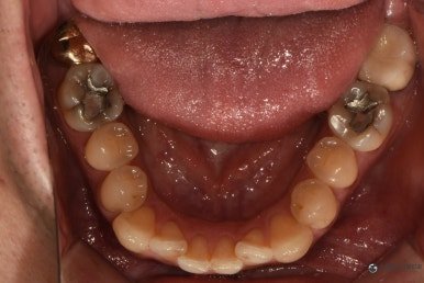
I Was Told the Molar With Root Canal Treatment Had to Be Extracted
The patient is a man in his 40s who lived in Germany.
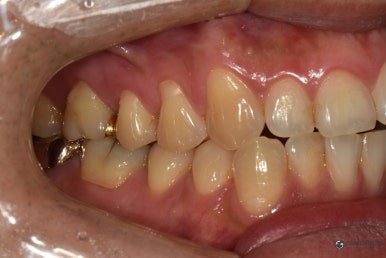
The last molar on the lower right had received root canal treatment at another clinic long ago
and had been restored with a gold crown.
Afterward he had discomfort around that tooth, and in Germany he was told about extracting it.
The patient came to the clinic wanting to check whether there was another treatment option
that could keep the tooth, if possible.
On checking the X-rays, around the roots of the molar that had previously had root canal treatment,
a relatively wide radiolucent area was seen.
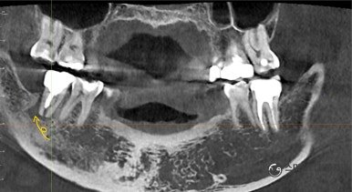
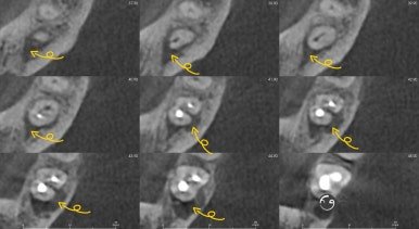
CT image before treatment
We Checked the Area Around the Root With CT
To check the area around the root in three dimensions, we also took a CT scan.
The CT cross-sections also showed a lesion around the root of that tooth
accompanied by loss of alveolar bone.
However, just because a lesion looks large on an X-ray,
this alone does not decide whether the tooth is extracted.
We have to evaluate together how the previous root canal treatment was done, whether there is any possibility of a crack or fracture in the root,
what the remaining tooth structure and periodontal tissue are like, whether the tooth can be restored again,
and so on.
When we checked the condition of this patient's previous root canal treatment, there was an area where the root canal filling seemed not to reach the root tip sufficiently.
After explaining the current condition and treatment options to the patient, before extraction,
we decided to try non-surgical root canal retreatment, that is, root canal retreatment first..
How Is Root Canal Retreatment Done?
Root canal retreatment is the process of treating again the inside of the root canal
of a tooth that has already had root canal treatment once.
Because the root canal filling material used in the previous treatment is still inside the canal,
we have to remove it and check the length and shape of the canal again.
For this patient as well, we used endodontic files to remove the previous root canal filling material
and check the condition inside the canal.
In retreatment, besides removing the old material, it is important to recheck
the path and working length of the previously treated canal, and
to secure an environment in which the inside of the canal can be cleaned and disinfected.
When the previous root canal filling did not reach the root tip sufficiently,
there can be various causes such as the shape of the canal or calcification,
so careful checking is needed during retreatment as well.
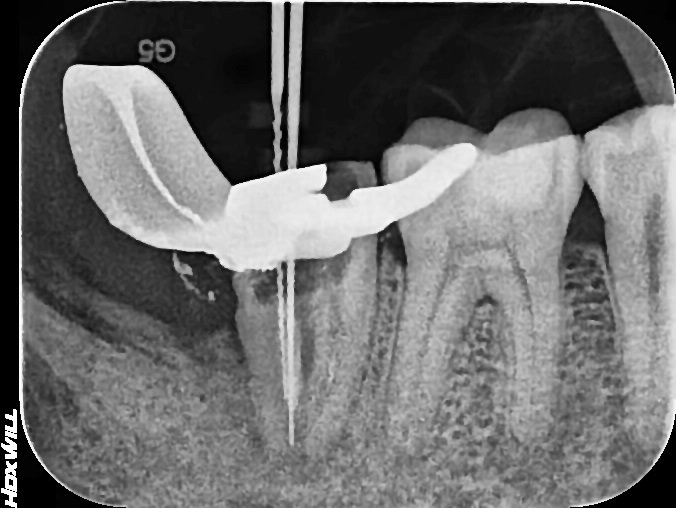
X-ray checking the canal length with a file
We Confirmed the Canal Path All the Way to the Root Tip.
After removing the previous root canal filling material, we rechecked the canal path and working length.
In this case, we were able to confirm the canal path toward the root tip within the treatable range,
and after cleaning and disinfecting the inside of the canal, we filled the canal again.
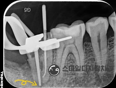
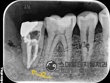
X-ray after treatment
Comparing the X-rays before and after treatment, you can see that after retreatment the root canal filling material extends
further toward the root tip than before treatment.
After completing the root canal retreatment, we finally proceeded with a crown, considering the remaining tooth structure and the bite.
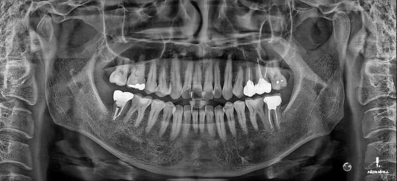
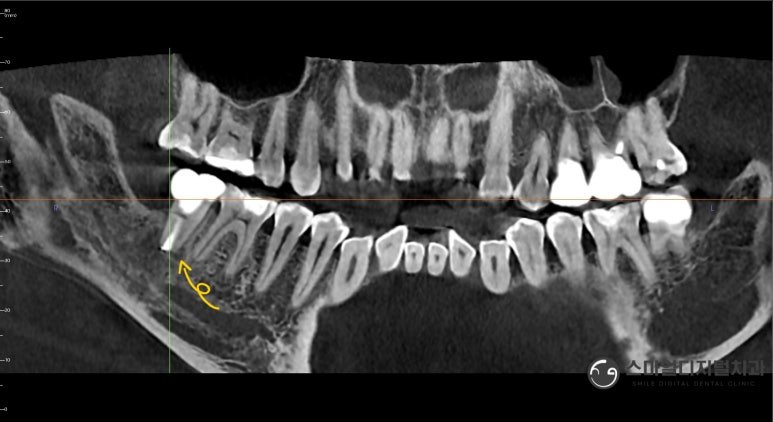
Panoramic and CT comparison after 6 months
6 Months Later, We Checked the Changes Around the Root
What matters in this case is the course after treatment.
Because a relatively wide radiolucent area was seen around the root before treatment,
we did not judge from the photos taken right after retreatment, but followed the progress over a period of time.
About 6 months after root canal retreatment, we took a panoramic X-ray and a CT scan.
Compared with before treatment, the radiolucent area that had been seen around the root decreased, and
signs of the surrounding bone tissue recoveringcould be seen.
However, the CT at that time still showed a radiolucent area around the root tip,
so we continued the follow-up.
On a panoramic X-ray alone, it may look as if a considerable change has occurred,
but because CT lets us check the area around the root more three-dimensionally,
we compared the two exams together.
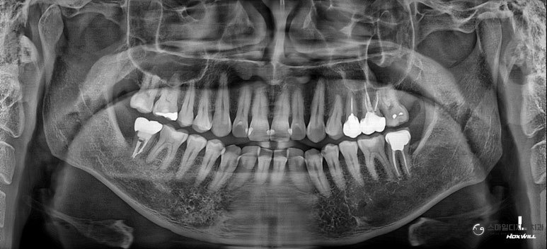
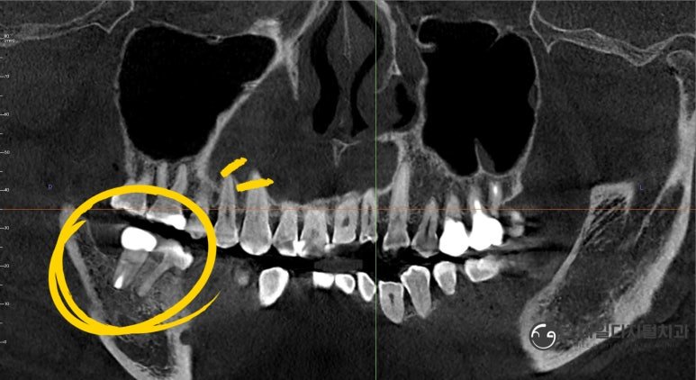
CT comparison at 1 year
Checking Again After 1 Year
About 1 year later, we checked the condition of the tooth again with a panoramic X-ray and CT.
Compared with before treatment, the radiolucent area around the root that had been seen earlier decreased further, and we could see the surrounding bone tissue recovering.
For a tooth that had a relatively wide lesion around the root like this,
it is important to observe the radiographic changes over a period of time rather than just the appearance right after treatment.
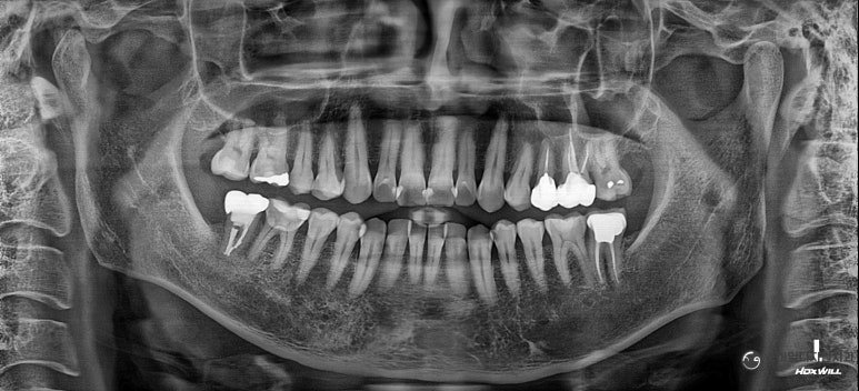
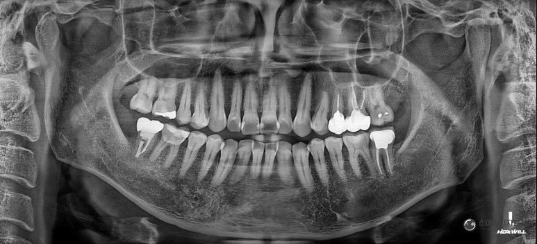
Panoramic X-rays at 2 years / 4 years
We Also Checked the Progress at 2 and 4 Years
After that, whenever the patient came to Korea, we checked the condition of that tooth.
In the X-rays taken about 2 years and 4 years after root canal retreatment, we again checked the treated tooth and
the condition of the surrounding bone tissue.
The wide radiolucent area around the root that had been seen before treatment decreased during follow-up after root canal retreatment,
and in the X-rays taken afterward, we continued to observe whether
the changed condition of the surrounding bone tissue after treatment was maintained.
In this case, rather than judging root canal retreatment as a success in one sentence, we think it is more important data to directly compare X-rays taken at several points in time, starting from before treatment.
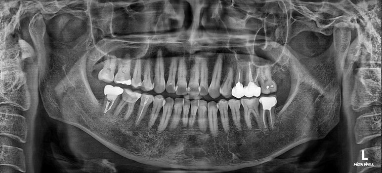
Most recent panoramic X-ray on October 2, 2026
October 2, 2026: A Checkup After His Recent Return to Korea.
The patient, who lives in Germany, recently came to Korea again,
and visited Smile Digital Dental Clinic on October 2, 2026 for a checkup.
This time too, we took a new panoramic X-ray and checked again the condition of the tooth that had undergone root canal retreatment and the surrounding tissue.
It was not the case that we checked only right after treatment and stopped there;
6 months → 1 year → 2 years → 4 years → most recent checkup on October 2, 2026
we have followed the changes in the same tooth over a long period, and that point
is the meaningful part of this case.
“At the most recent checkup as well, the patient did not complain of any particular discomfort
related to that tooth.”
If a Tooth With Previous Root Canal Treatment Has a Problem Again, Can All Be Retreated?
Not necessarily.
Even if there is a lesion around the roots of a tooth that previously had root canal treatment,
not every tooth can be kept through root canal retreatment.
Conversely, just because a lesion is seen around the root,
not every such tooth immediately has to be extracted.
We need to evaluate together the condition of the previous root canal treatment, the shape of the canals, whether the root is cracked or fractured,
the amount of remaining natural tooth, the condition of the periodontal tissue, and finally
whether the tooth can be restored again.
Depending on the condition, non-surgical root canal retreatment may be considered,
in some cases other methods such as apical surgery may be considered,
and if it is judged that the tooth is difficult to keep, extraction may be necessary.
Therefore, rather than regarding root canal retreatment as the last treatment before extraction,
it is better to understand it as one of the treatment options that can be considered after evaluating whether
there is a possibility of preserving the current tooth.
Root Canal Retreatment Also Needs Follow-Up After Treatment.
With root canal retreatment, the whole process is not over just because the root canal treatment is finished.
In particular, when a relatively wide lesion was seen around the root before treatment, as in this patient,
it is important to check how the tissue around the root changes through X-ray exams over a period of time,
along with changes in symptoms after treatment.
This patient's course was as follows:
discomfort in the tooth with previous root canal treatment → panoramic X-ray and CT → checking the condition of the previous root canal treatment → root canal retreatment → final crown → 6 months → 1 year → 2 years → 4 years
→ most recent checkup on October 2, 2026,
and treatment and follow-up continued in this order.
At first a relatively wide lesion was seen around the root, but
rather than deciding right away whether to extract, we evaluated the current tooth and the condition of the previous root canal treatment,
carried out root canal retreatment, and afterward followed the changes in the surrounding tissue
through X-rays over a long period.
If a problem has come up again in a tooth that previously had root canal treatment,
rather than judging that it can definitely be saved or that it definitely has to be extracted,
it is important to first evaluate the current condition of the tooth and the possibility of retreatment.
The photos of the treatment process and follow-up in this case are published with the patient's consent.
Treatment methods and results vary from person to person. Depending on the condition of the previous root canal treatment, the shape of the canals, whether the root is cracked or fractured, the periodontal tissue, and the remaining tooth structure, root canal retreatment may be difficult or the tooth may not be preserved. Symptoms may continue after treatment, and additional root canal treatment, surgical treatment, or extraction may be needed. The specific treatment method is decided after examination and diagnosis.
Related cases:
Front Tooth Crown Retreatment in Busan Seomyeon: A Case With Root Canal Retreatment and Non-Vital Whitening
Front Tooth Crown in Seomyeon, Busan: Non-Vital Tooth Whitening and Gum Surgery Together
Seomyeon, Busan Front Tooth Crown: Re-Treating a Root-Only Tooth While Preserving It
Related information: Same-day crown & one-day restoration guide · General dental information is also available on the Korean Dental Association.